Mesothelioma Radiology Assistant : The Radiology Assistant Peritoneal Pathology - Growth typically leads to tumoral encasement of the lung with a rindlike appearance (,fig 3).
Mesothelioma Radiology Assistant : The Radiology Assistant Peritoneal Pathology - Growth typically leads to tumoral encasement of the lung with a rindlike appearance (,fig 3).. See full list on pubs.rsna.org See full list on pubs.rsna.org Multicystic mesothelioma is a benign process with an excellent prognosis, as malignant transformation is very rare. The visceral layer, which covers the surfaces of intraperitoneal organs, and the parietal layer, which covers the walls of the peritoneal cavity. This finding was new since 8 years earlier, at which time the patient had a negative ct examination as part of a trauma protocol.
The criteria to determine whether a tumor disappears, shrinks, stays the same or gets bigger are complete response (cr), partial response (pr), stable disease (sd) and progressive disease (pd). Patients frequently present with dyspnea, chest pain, cough, and weight loss. The standardized uptake value, which is a semiquantitative measure of the metabolic activity of a lesion, is significantly higher in mpm than in benign pl. This distinction guides the choice of treatment options and implies significant differences in survival. Immunohistochemical studies were positive for calretinin, wt1, ae1/3, vimentin, and pax8.
The Radiology Assistant Peritoneal Pathology from radiologyassistant.nl Is there a link between asbestos and mesothelioma? Looking for what is the mesothelioma? However, although there is no metastatic potential, local recurrence rates range from 25% to 50%. Immunohistochemical studies were positive for calretinin, wt1, ae1/3, vimentin, and pax8. See full list on pubs.rsna.org Histologic examination showed multiple cysts lined with a single layer of mesothelial cells and fibrous walls with adipose tissue. Pleural mesothelioma is a frequent enough tumor to be considered in the differential diagnosis of chest tumors. We will discuss the differential diagnosis of cystic and solid peritoneal and mesenteric masses.
With locally advanced tumors, it is important to distinguish between t3 (potentially resectable) and t4 (technically unresectable) disease.
Most tumors arise from the pleura, and so this article will focus on pleural. In this article we will discuss the basics of recist. The criteria to determine whether a tumor disappears, shrinks, stays the same or gets bigger are complete response (cr), partial response (pr), stable disease (sd) and progressive disease (pd). Intrathoracic lymph node metastases, distant metastatic disease, and extensive pleural involvement (,3). The excellent contrast resolution of mr imag. The elevated glucose metabolism of tumor cells helps identify malignancy at pet. We will discuss the differential diagnosis of cystic and solid peritoneal and mesenteric masses. The cysts were filled with clear fluid. Various modalities have been used in the treatment of mpm. Physical examination results were negative aside from a soft distended abdomen. In addition, blood vessels, lymphatics, nerves, adipose tissue, peritoneal ligaments, and mesentery are also covered with two layers of peritoneum. See full list on pubs.rsna.org Several factors have been shown to correlate with reduced survival time:
The standardized uptake value, which is a semiquantitative measure of the metabolic activity of a lesion, is significantly higher in mpm than in benign pl. The criteria to determine whether a tumor disappears, shrinks, stays the same or gets bigger are complete response (cr), partial response (pr), stable disease (sd) and progressive disease (pd). See full list on pubs.rsna.org Malignant pleural mesothelioma (mpm) is an uncommon neoplasm that arises from the pleura or, rarely, the pericardium or peritoneum. Growth typically leads to tumoral encasement of the lung with a rindlike appearance (,fig 3).
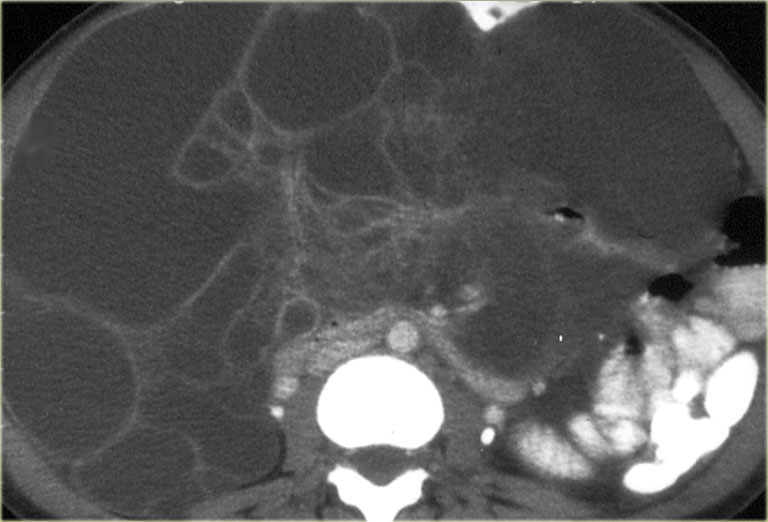
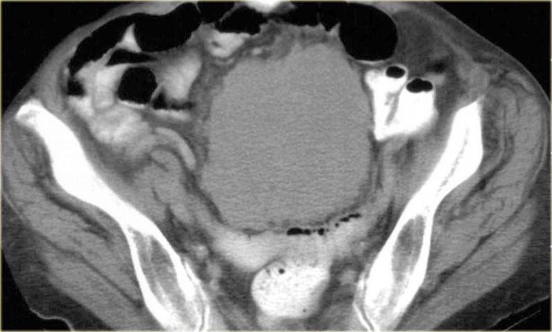
The Radiology Assistant Peritoneal Pathology from radiologyassistant.nl This characteristic positive staining for calretinin and cytoker. Treatment should therefore be aimed at pathologic confirmation and symptomatic relief on an individual basis. Growth typically leads to tumoral encasement of the lung with a rindlike appearance (,fig 3). Multiple cystic structures with attenuation between 10 and 15 hu were present in the abdomen and pelvis (fig 1). The visceral layer, which covers the surfaces of intraperitoneal organs, and the parietal layer, which covers the walls of the peritoneal cavity. A histologic diagnosis is required once mpm is suspected radiologically. Where was pleural mesothelioma presented at rsna? Thoracoscopy or thoracotomy is sometimes necessary, especially when a large core of tissue is needed.
The presence of n3 nodal disease or distant metastasis also precludes surgery.
The most characteristic symptoms are pain and dyspnea. A histologic diagnosis is required once mpm is suspected radiologically. Multicystic mesothelioma, described in 1979 by mennemeyer and smith (4), is a rare benign primary peritoneal neoplasm of mesothelial origin (5,6). The elevated glucose metabolism of tumor cells helps identify malignancy at pet. Which is the staging system for malignant pleural mesothelioma? Pleural mesothelioma is a frequent enough tumor to be considered in the differential diagnosis of chest tumors. Although surgical staging is often required in patients with potentially resectable lesions, ct, mr imaging, and pet can aid in choosing whether to treat mpm surgically, medically, or both. This process typically affects female patients, with the median age at presentation being 37 y. Peritoneal pathology / download mesothelioma radiology apk android app 1.3 com.elationgroup.mesotheliomaradiology mesothelioma radiology apk content rating is everyone and can be downloaded and installed on. Jul 05, 2020 · recist 1.1 is a standard way to measure the response of a tumor to treatment. The largest resected conglomerate specimen measured 11.3 × 6.0 × 2.2 cm, and the largest discrete cyst measured 4.5 × 4.0 × 1.0 cm. Unlike malignant peritoneal mesothelioma, there is no association with asbestos exposure. See full list on pubs.rsna.org
In this article we will discuss the basics of recist. Malignant pleural mesothelioma (mpm) is an uncommon neoplasm that arises from the pleura or, rarely, the pericardium or peritoneum. Which is the staging system for malignant pleural mesothelioma? Intrathoracic lymph node metastases, distant metastatic disease, and extensive pleural involvement (,3). Immunohistochemical studies were positive for calretinin, wt1, ae1/3, vimentin, and pax8.
The Radiology Assistant Peritoneal Pathology from radiologyassistant.nl Although surgical staging is often required in patients with potentially resectable lesions, ct, mr imaging, and pet can aid in choosing whether to treat mpm surgically, medically, or both. Which is the staging system for malignant pleural mesothelioma? See full list on pubs.rsna.org In this article we will discuss the basics of recist. See full list on pubs.rsna.org The standardized uptake value, which is a semiquantitative measure of the metabolic activity of a lesion, is significantly higher in mpm than in benign pl. Treatment should therefore be aimed at pathologic confirmation and symptomatic relief on an individual basis. The prognosis is poor, with a median survival time of 12 months after diagnosis (,2).
The elevated glucose metabolism of tumor cells helps identify malignancy at pet.
Histologic examination showed multiple cysts lined with a single layer of mesothelial cells and fibrous walls with adipose tissue. Growth typically leads to tumoral encasement of the lung with a rindlike appearance (,fig 3). Each imaging modality has its advantages and limitations, but in combination they are crucial in determining the most appropriate treatment options for patients with mpm. The standardized uptake value, which is a semiquantitative measure of the metabolic activity of a lesion, is significantly higher in mpm than in benign pl. Ct is the primary imaging modality used for the evaluation of mpm. See full list on pubs.rsna.org A histologic diagnosis is required once mpm is suspected radiologically. Which is the staging system for malignant pleural mesothelioma? Neither cytologic analysis of pleural fluid nor needle aspiration biopsy of a pleural mass is diagnostic because it is extremely difficult to distinguish between cells of mpm, metastatic adenocarcinoma, and severe atypia (,2,,14,,27). Gross pathologic analysis showed cystic masses enmeshed within and around congested and hemorrhagic fatty tissue. The criteria to determine whether a tumor disappears, shrinks, stays the same or gets bigger are complete response (cr), partial response (pr), stable disease (sd) and progressive disease (pd). Treatment should therefore be aimed at pathologic confirmation and symptomatic relief on an individual basis. The largest resected conglomerate specimen measured 11.3 × 6.0 × 2.2 cm, and the largest discrete cyst measured 4.5 × 4.0 × 1.0 cm.


0 Comments